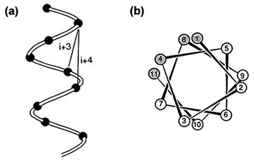
Amphipathic α-helices can be found at the surface of a water-soluble globular protein, whereas hydrophobic helices are on the inside. As the name implies, an amphipathic (or amphiphilic) helix is an α-helix with both hydrophobic and hydrophilic amino acid residues arranged in such a way as to create two faces on opposite sides of the helix, one face being hydrophobic.
When soluble proteins fold in water, the most stable conformation ought to be the one in which the maximum number of polar groups is on the surface and in contact with water, while at the same time the maximum number of nonpolar side chains is buried and away from the surface. This means that nonpolar side chains in an α-helix near the surface will be on one side of the helix, whereas the polar side chains will tend to be on the other side, where they can project into the water phase. Thus, the amino acid sequence of an amphiphilic helix is quite characteristic; amino acids that are definitely hydrophilic occur at every second or third position in the sequence. At the same time, strongly hydrophobic side chains will be found at every third or fourth position. This spacing is illustrated in .
The cartoon model of barnase, an extracellular 110-residue ribonuclease from Bacillus amyloliquefaciens. Note that all three α-helices are at the surface.
the α-helix (residues 7-17). The side chains are omitted for clarity.
Click to view the α-helix end-on.
Note how the hydrophobic side chains of Phe-7, Val-10, and Leu-14, and the hydrophobic part of Tyr-13 are on one side of the helix. This side packs against hydrophobic side chains in the nonpolar interior of the protein. The other side of the helix, which is exposed to the solvent water, contains hydrophilic side chains. This is a typical amphipathic helix.
Space-filling model of barnase. The hydrophobic side chains of the α-helix are shown in magenta; the hydrophilic side chains of the helix are shown in purple.